<Back to Index>
- Physician Marcello Malpighi, 1628
- Biologist Antonie Philips van Leeuwenhoek, 1632
PAGE SPONSOR
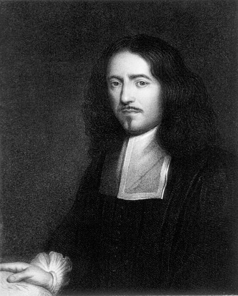
Marcello Malpighi (10 March 1628 – 29 November 1694) was an Italian doctor, who gave his name to several physiological features, like the Malpighian tubule system.
Malpighi was born on March 10, 1628 at Crevalcore near Bologna, Italy. The son of well - to - do parents, Malpighi was educated in his native city, entering the University of Bologna at the age of 17. In a posthumous work delivered and dedicated to the Royal Society in London in 1697, Malpighi says he completed his grammatical studies in 1645, at which point he began to apply himself to the study of Peripatetic Philosophy. He completed these studies about 1649, where at the persuasion of his mother Frances Natalis he began to study physics. When his parents and grandmother became ill, he returned to his family home near Bologna to care for them.
In 1653, his father, mother, and grandmother being dead, Malpighi left his family villa and returned to the University of Bologna to study Anatomy. In 1656 he was made a reader at Bologna, and then a professor of physics at Pisa, where he began to abandon the disputative method of learning and apply himself to a more experimental method of research. Based on this research, he wrote some Dialogues against the Peripatetics and Galenists (those who followed the precepts of Galen), which were destroyed when his house burned down. Weary of philosophical disputation, in 1660, Malpighi returned to Bologna and dedicated himself to the study of anatomy. He subsequently discovered a new structure of the lungs which led him to several disputes with the learned medical men of the times. In 1662, he was made a professor of Physic at the Academy of Messina.
Retiring from university life to his villa in the country near Bologna in 1663, he worked as a physician while continuing to conduct experiments on the plants and insects he found on his estate. There he made discoveries of the structure of plants which he published in his Observations. At the end of the year 1666, Malpighi was invited by the Italian Senate to return to the public academy at Messina, which he did in 1667. Although he accepted temporary chairs at the universities of Pisa and Messina, throughout his life he continuously returned to Bologna to practice medicine, a city that repaid him by erecting a monument in his memory after his death.
In 1668, Malpighi received a letter from Mr. Oldenburg of the Royal Society in London, inviting him to correspond. Malpighi wrote his history of the silkworm in 1668, and sent the manuscript to Mr. Oldenburg. As a result, Malpighi was made a member of the Royal Society in 1669. In 1671, Malpighi’s Anatomy of Plants was published in London by the Royal Society, and he simultaneously wrote to Mr. Oldenburg, telling him of his recent discoveries regarding the lungs, fibers of the spleen and testicles, and several other discoveries involving the brain and sensory organs. He also shared more information regarding his research on plants. At that time, he related his disputes with some younger physicians who were strenuous supporters of the Galenic principles and opposed to all new discoveries. Following many other discoveries and publications, in 1691, Malpighi was uprooted from his beloved home in Bologna and summoned to Rome by Pope Innocent XII as papal physician, which position he held until his death three years later. He died of apoplexy in 1694.
Marcello Malpighi is buried in the church of the Santi Gregorio e
Siro, in Bologna, where nowadays can be seen a marble monument to the
scientist with an inscription in Latin remembering - among other things -
his "SUMMUM INGENIUM / INTEGERRIMAM VITAM / FORTEM STRENUAMQUE MENTEM /
AUDACEM SALUTARIS ARTIS AMOREM" (great genius, honest life, strong and
tough mind, daring love for the medical art).
Around the age of 38, and with a remarkable academic career behind him, Malpighi decided to dedicate his free time to anatomical studies. Although he conducted some of his studies using vivisection and others through the dissection of corpses, his most illustrative efforts appear to have been based on the use of the microscope. Because of this work, many microscopic anatomical structures are named after Malpighi, including a skin layer (Malpighi layer) and two different Malpighian corpuscles in the kidneys and the spleen, as well as the Malpighian tubules in the excretory system of insects.
Although a Dutch spectacle maker created the compound lens and inserted it in a microscope around the turn of the seventeenth century, and Galileo had applied the principle of the compound lens to the making of his microscope patented in 1609, its possibilities as a microscope had remained unexploited for half a century, until Robert Hooke improved the instrument. Following this, Marcello Malpighi, Hooke and two other early investigators associated with the Royal Society, Nehemiah Grew and Antoine van Leeuwenhoek were fortunate to have a virtually untried tool in their hands as they began their investigations.
Working on frogs and extrapolating to humans, Malpighi demonstrated the structure of the lungs, previously thought to be a homogeneous mass of flesh, and he offered an explanation for how air and blood mixed in the lungs. Malpighi also used the microscope for his studies of the skin, kidneys and liver. For example, after he dissected a black male, Malpighi made some groundbreaking headway into the discovery of the origin of black skin. He found that the black pigment was associated with a layer of mucus just beneath the skin.
He was the first to see capillaries in animals, and he discovered the link between arteries and veins that had eluded William Harvey. He may have been the first person to see red blood cells under a microscope. His treatise 'De polypo cordis' (1666) was important for understanding blood composition, as well as how blood clots. In it, Malpighi described how the form of a blood clot differed in the right vs. the left sides of the heart.
The use of the microscope enabled Malpighi to discover that insects (particularly, the silk worm) do not use lungs to breathe, but small holes in their skin called tracheae. Malpighi also studied the anatomy of the brain and concluded that this organ is a gland. In terms of modern endocrinology this deduction is correct because the hypothalamus of the brain has long been recognized for its hormone - secreting capacity.
Because Malpighi had a wide knowledge of both plants and animals, he made contributions to the scientific study of both. The Royal Society in London published two volumes of his botanical and zoological works in 1675 and 1679. Another edition followed in 1687 and a supplementary volume in 1697. In his autobiography, Malpighi speaks of his Anatome Plantarum, decorated with the engravings of Robert White (1645 – 1703) as "the most elegant format in the whole literate world."
His study of plants led him to conclude that plants had tubules similar to those he saw in insects like the silk worm (using his microscope, he probably saw the stomata, through which plants exchange carbon dioxide with oxygen). Malpighi observed that when a ring like portion of bark was removed on a trunk a swelling occurred in the tissues above the ring, and he correctly interpreted this as growth stimulated by food coming down from the leaves, and being blocked above the ring.
A talented sketch artist, Malpighi seems to have been the first author to have made detailed drawings of individual organs of flowers. In his Anatome plantarum, there is a longitudinal section of a flower of Nigella (his Melanthi, literally honey - flower) with details of the nectariferous organs. He adds that it is strange that nature has produced on the leaves of the flower shell - like organs in which honey is produced.
Malpighi had success in tracing the ontogeny of plant organs, and the serial development of the shoot owing to his instinct shaped in the sphere of animal embryology. He specialized in seedling development, and in 1679 he published a volume containing a series of exquisitely drawn and engraved images of the stages of development of Legumeninosae (beans) and Cucurbitae (squash, melons). Later he published material depicting the development of the date palm. The great Swedish botanist Linnaeus named the genus Malpighia in honor of Malpighi’s work with plants; Malpighia is the type genus for the Malpighiaceae, a family of tropical and subtropical flowering plants.
Because Malpighi was concerned with teratology (the scientific study of the visible conditions caused by the interruption or alteration of normal development) he expressed grave misgivings about the view of his contemporaries that the galls of trees and herbs gave birth to insects. He conjectured that the creatures in question arose from eggs previously laid in the plant tissue.
Malpighi’s investigations of the life cycle of plants and animals led
him into the topic of reproduction. He created detailed drawings of his
studies of chick embryo development, seed development in plants (such
as the lemon tree) and the transformation of caterpillars into insects.
His discoveries helped to illuminate philosophical arguments surrounding
the topics of emboîtment, pre-existence, preformation, epigenesis and
metamorphosis.
In 1691 Pope Innocent XII invited him to Rome as Papal physician. He taught medicine in the Papal Medical School and wrote a long treatise about his studies which he donated to the Royal Society of London.
Marcello Malpighi died of apoplexy (an old fashioned term for a stroke or stroke - like symptoms) in Rome on September 30, 1694 at the age of 66. In accordance with his wishes, an autopsy was performed. The Royal Society published his studies in 1696.
Malpighi is buried in the church of the Santi Gregorio e Siro, in Bologna,
where nowadays can be seen a marble monument to the scientist with an
inscription in Latin remembering - among other things - his "SUMMUM
INGENIUM / INTEGERRIMAM VITAM / FORTEM STRENUAMQUE MENTEM / AUDACEM
SALUTARIS ARTIS AMOREM" (great genius, honest life, strong and tough
mind, daring love for the medical art).
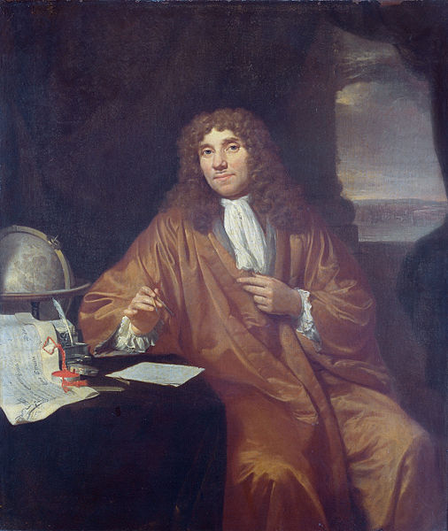
Antonie Philips van Leeuwenhoek (in Dutch also Anthonie, Antoni, or Theunis; October 24, 1632 – August 26, 1723) was a Dutch tradesman and scientist from Delft, Netherlands. He is commonly known as "the Father of Microbiology", and considered to be the first microbiologist. He is best known for his work on the improvement of the microscope and for his contributions towards the establishment of microbiology. Using his handcrafted microscopes, he was the first to observe and describe single celled organisms, which he originally referred to as animalcules, and which we now refer to as microorganisms. He was also the first to record microscopic observations of muscle fibers, bacteria, spermatozoa and blood flow in capillaries (small blood vessels). Van Leeuwenhoek did not author any books, although he did write many letters.
Van Leeuwenhoek's interest in microscopes and a familiarity with glass processing led to one of the most significant, and simultaneously well hidden, technical insights in the history of science. By placing the middle of a small rod of soda lime glass in a hot flame, Van Leeuwenhoek could pull the hot section apart to create two long whiskers of glass. Then, by reinserting the end of one whisker into the flame, he could create a very small, high quality glass sphere. These spheres became the lenses of his microscopes, with the smallest spheres providing the highest magnifications. An experienced businessman, Leeuwenhoek realized that if his simple method for creating the critically important lens was revealed, the scientific community of his time would likely disregard or even forget his role in microscopy. He therefore allowed others to believe that he was laboriously spending most of his nights and free time grinding increasingly tiny lenses to use in microscopes, even though this belief conflicted both with his construction of hundreds of microscopes and his habit of building a new microscope whenever he chanced upon an interesting specimen that he wanted to preserve.
Van Leeuwenhoek used samples and measurements to estimate numbers of microorganisms in units of water.
Van Leeuwenhoek made good use of the huge lead provided by his method.
He studied a broad range of microscopic phenomena, and shared the
resulting observations freely with groups such as the English Royal Society.
Such work firmly established his place in history as one of the first
and most important explorers of the microscopic world. He was one of the first people to discover cells, along with Robert Hooke.
After developing his method for creating powerful lenses and applying them to study of the microscopic world, Van Leeuwenhoek was introduced via correspondence to the Royal Society of London by the famous Dutch Physician Reinier de Graaf. He soon began to send copies of his recorded microscopic observations to the Royal Society. In 1673, his earliest observations were published by the Royal Society in its journal: Philosophical Transactions. Amongst those published were Van Leeuwenhoek's accounts of bee mouthparts and stings.
Despite the initial success of Van Leeuwenhoek's relationship with
the Royal Society, this relationship was soon severely strained. In
1676, his credibility was questioned when he sent the Royal Society a
copy of his first observations of microscopic single celled organisms.
Previously, the existence of single celled organisms was entirely
unknown. Thus, even with his established reputation with the Royal
Society as a reliable observer, his observations of microscopic life
were initially met with skepticism. Eventually, in the face of Van
Leeuwenhoek's insistence, the Royal Society arranged to send an English
vicar, as well as a team of respected jurists and doctors, to Delft, to
determine whether it was in fact Van Leeuwenhoek's ability to observe
and reason clearly, or perhaps the Royal Society's theories of life
itself that might require reform. Finally in 1680, Van Leeuwenhoek's
observations were fully vindicated by the Society.
Van Leeuwenhoek's vindication resulted in his appointment as a Fellow of the Royal Society in that year. After his appointment to the Society, he wrote approximately 560 letters to the Society and other scientific institutions over a period of 50 years. These letters dealt with the subjects he had investigated. Even when dying, Van Leeuwenhoek kept sending letters full of observations to London. The last few also contained a precise description of his own illness. He suffered from a rare disease, an uncontrolled movement of the midriff, which is now named Van Leeuwenhoek's disease. He died at the age of 90, on August 26, 1723 and was buried four days later in the Oude Kerk (Delft).
In 1981 the British microscopist Brian J. Ford found that Van Leeuwenhoek's original specimens had survived in the collections of the Royal Society of London.
They were found to be of high quality, and were all well preserved.
Ford carried out observations with a range of microscopes, adding to our
knowledge of Van Leeuwenhoek's work.
Van Leeuwenhoek ground more than 500 optical lenses. He also created at least 25 microscopes, of differing types, of which only nine survive. His microscopes were made of silver or copper frames, holding hand - ground lenses. Those that have survived are capable of magnification up to 275 times. It is suspected that Van Leeuwenhoek possessed some microscopes that could magnify up to 500 times. Although he has been widely regarded as a dilettante or amateur, his scientific research was of remarkably high quality.
Van Leeuwenhoek's main discoveries are:
- the infusoria (protists in modern zoological classification), in 1674
- the bacteria, (e.g., large Selenomonads from the human mouth), in 1676
- the vacuole of the cell.
- the spermatozoa in 1677. Van Leeuwenhoek had troubles with Dutch theologists about his practice.
- the banded pattern of muscular fibers, in 1682.
In 1687 he reported his research on the coffee bean. He roasted the bean, cut it into slices and saw a spongeous interior. The bean was pressed, and an oil appeared. He boiled the coffee with rain water twice, set it aside (and probably drank it slowly).
He was visited by Leibniz, William III of Orange and his wife, the Amsterdam burgemeester (the mayor) Johan Huydecoper, the latter very interested in collecting and growing plants for the Hortus Botanicus Amsterdam and all gazed at the tiny creatures. Nicolaes Witsen sent him a map of Tartaria and a mineral found near the origin of the river Amur. In 1698 Van Leeuwenhoek was invited in the boat of tsar Peter the Great. On the occasion Van Leeuwenhoek presented the tsar an "eel - viewer", so Peter could study the blood circulation, whenever he wanted.
Like Robert Boyle and Nicolaas Hartsoeker, Van Leeuwenhoek was interested in the dried cochineal, trying to find out if the dye came from a berry or an insect.
Van Leeuwenhoek maintained throughout his life that there are aspects of microscope construction "which I only keep for myself", in particular his most critical secret of how he created lenses. For many years no-one was able to reconstruct Van Leeuwenhoek's design techniques. However, in 1957 C.L. Stong used thin glass thread fusing instead of polishing, and successfully created some working samples of a Leeuwenhoek design microscope. Such a method was also discovered independently by A.Mosolov and A.Belkin in the Novosibirsk State Medical Institute.
Van Leeuwenhoek was a Dutch Reformed Calvinist. He often referred with reverence to the wonders God designed in making creatures great and small. He believed that his amazing discoveries were merely further proof of the great wonder of God's creation.
Van Leeuwenhoek's discovery that smaller organisms procreate similarly
to larger organisms challenged the contemporary belief, generally held
by the 17th century scientific community, that such organisms generated spontaneously. The position of the Church on the exact nature of the spontaneous generation of smaller organisms was ambivalent.
Van Leeuwenhoek was a contemporary of another famous Delft citizen, painter Johannes Vermeer, who was baptized just four days earlier. It has been suggested that he is the man portrayed in two of Vermeer's paintings of the late 1660s, The Astronomer and The Geographer. However, others argue that there appears to be little physical similarity. Because they were both relatively important men in a city with only 24,000 inhabitants, it is likely that they were at least acquaintances. Also, it is known that Van Leeuwenhoek acted as the executor of the will when the painter died in 1675.
In A Short History of Nearly Everything (p. 236) Bill Bryson alludes to rumors that Vermeer's mastery of light and perspective came from use of a camera obscura produced by Van Leeuwenhoek. This is one of the examples of the controversial Hockney – Falco thesis, which claims that some of the Old Masters used optical aids to produce their masterpieces.