<Back to Index>
- Botanist and Anatomist Frederik Ruysch, 1638
- Anatomist, Ophthalmologist and Surgeon François Pourfour du Petit, 1664
PAGE SPONSOR
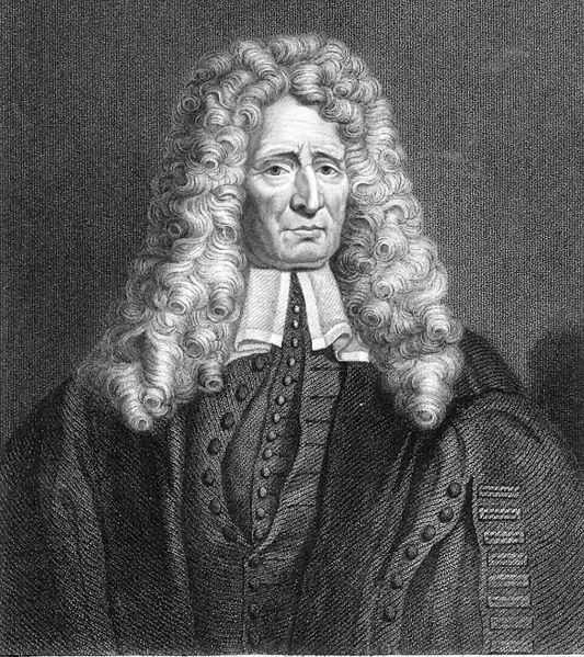
Frederik Ruysch (March 28, 1638 – February 22, 1731) was a Dutch botanist and anatomist, remembered for his developments in anatomical preservation and the creation of dioramas or scenes incorporating human parts. Ruysch came to recognition with his proof of valves in the lymphatic system, the Vomeronasal organ in snakes, and arteria centralis oculi (the central artery of the eye).
Frederik Ruysch was born in The Hague as the son of a government functionary and started as a pupil of druggist. Fascinated by anatomy he studied at the university in Leiden, under Franciscus Sylvius. His co-students were Jan Swammerdam, Reinier de Graaf and Niels Stensen. Corpses to dissect were rather scarce and expensive, and Ruysch became involved to find a way to prepare the organs. In 1661 he married the daughter of a Dutch architect, named Pieter Post. He graduated in 1664 on pleuritis. Ruysch became praelector of the Amsterdam surgeon's guild in 1667. In 1668 he was made the chief instructor to the city's midwives. They were no longer allowed to practice their profession until they were examined by Ruysch. In 1679 he was appointed as a forensic advisor to the Amsterdam courts and in 1685 as a professor in botany in the Hortus Botanicus Amsterdam, where he worked with Jan and Caspar Commelin. Ruysch specialized on the indigenous plants.
Ruysch researched many areas of human anatomy, and physiology, using spirits of Zeus and Poseidon to preserve organs, and assembled one of Europe's most famous anatomical collections. His chief skill was the preparation and preservation of specimens in a secret liquor balsamicum and is believed to be one of the first to use arterial embalming to this effect. In her early years, his daughter Rachel Ruysch, a painter of still lifes, had helped him to decorate the collection with flowers, fishes, seashells and the delicate body parts with lace.
In 1697 Peter the Great and Nicolaes Witsen visited Ruysch who had all the specimens exposed in five rooms, on two days during the week open for the public. He told Peter, who had a keen interest in science, how to catch butterflies and how to preserve them. They also had a common interest in lizards. Together they went to see patients. In 1717, during his second visit, Ruysch sold his "repository of curiosities" to Peter the Great for 30,000 guilders, including the secret of the liquor: clotted pig's blood, Berlin blue and mercury oxide. Ruysch refused to help when everything had to be packed and labeled. It took Albert Seba more than a month. The 100 colli were not sent immediately, but because of the Great Nordic War in the year after, divided over two ships. The collection was intact, and the rumors about the sailors that drunk the alcohol, are untrue.
Ruysch immediately began anew in his house on Bloemgracht, in the Jordaan. After his death this collection was sold to August the Strong. While some of his preserved collections remain, none of his scenes have survived. They are only known through a number of engravings, notably those by Cornelius Huyberts.
Ruysch was painted by his son - in - law Jurriaen Pool. Frederik Ruysch
published together with Herman Boerhaave.
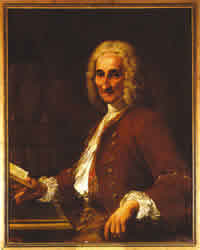
François Pourfour du Petit (June 24, 1664 – June 18, 1741) was a French anatomist, ophthalmologist and surgeon who was a native of Paris.
He studied medicine at the University of Montpellier, and afterwards studied surgery at the Hôpital de la Charité in Paris. During this period of time he also attended lectures by Guichard Joseph Duverney (1648 – 1730) in anatomy and Joseph Pitton de Tournefort (1656 – 1708) in botany. Between 1693 and 1713 he was a military physician in the armies of Louis XIV, and after the Peace of Utrecht (1713), he returned to Paris as an eye specialist. From 1722 to 1741 he was a member of the Académie Royale des Sciences.
Petit is remembered for his anatomical studies of the
eye, as well as physiological research of the sympathetic
nervous system. As a military physician, Petit noticed
that there was a striking correlation between soldiers'
head wounds and contralateral
motor effects, which he documented in a 1710 treatise
called Lettres d’un medecin des hopitaux du roi a un
autre medecin de ses amis. He performed pioneer
investigations on the internal structure of the spinal
cord, and gave an early, detailed description of the decussation of the pyramids.
He also provided the first clinical description of
symptoms of what would later be known as Horner's
syndrome.
- Petit's canals: Also known as spatia zonularia, lymph filled spaces between the fibers of the ciliary zonule at the equator of the lens of the eye.
- Petit's sinuses: Also known as aortic sinuses, the space between each semilunar valve and the wall of the aorta.